2026年8月7日,Sabira Alpysbayeva等,在《Small Ruminant Research》,发表“Comparison of subcutaneous and conjunctival Brucella melitensis Rev.1 vaccination in small ruminants: Serological response and post-challenge bacterial dissemination”的论文。
研究要点
布鲁氏菌(Brucella melitensis)Rev. 1 广泛用于控制绵羊和山羊的布鲁氏菌病,但其接种途径可能影响保护效果及接种后的血清学干扰。
将 25 只本地品种绵羊和山羊分别分配至皮下接种组(SC,n = 10)、结膜接种组(VC,n = 10)以及未接种但接受攻毒的对照组(n = 5)。
各接种组在接种途径、剂量及辅料方面均存在差异(SC:2 × 10⁹ CFU 溶于 2 mL 常规稀释液;VC:1 × 10⁹ CFU 溶于 40 µL 含 5% Montanide Gel-01 的稀释液),因此比较结果反映了这三个因素的综合作用,而非仅接种途径的影响。
通过间接 ELISA 试验和玫瑰红试验,对血清学结果进行了为期 15 个月的监测,之后所有动物均接受强毒力布鲁氏菌 16 M 的结膜攻毒,并于 15 天后实施安乐死。
皮下接种可诱导持续抗体反应,第 15 个月时抗体水平仍高于基线值(S/P % = 181.4 ± 49.4),且 4/10 只动物仍呈玫瑰红阳性。
结膜接种则仅引发短暂抗体反应,抗体水平始终未达阳性阈值(最高为 36.1%),且所有 VC 组动物从第 6 个月起均呈玫瑰红阴性(第 6 个月:10/10 vs 0/10;Holm 校正后 p = 6.5 × 10⁻⁵)。
攻毒后,从10只SC组、4/10只VC组及5/5只对照组动物中分别分离到布鲁氏菌;
两个疫苗接种组间的差异无统计学意义(p = 0.17)。
VC组的细菌载量显著较低(中位数:0 vs. 每只动物262.5个菌落;p = 0.0052),且VC组中布鲁氏菌的分离仅见于淋巴结,而SC组动物亦可从脾脏及肝脏中分离到该菌。
所有分离株均鉴定为16M株。
两种接种途径均未诱导无菌免疫。
在本小规模队列及存在混杂因素的研究设计限制下,结膜给药方案既能降低血清学持续阳性率,又可限制细菌播散范围——这一组合策略对于需在维持血清学监测兼容性的前提下进行疫苗接种的场景可能更具优势。
图文速览
Table 1 Demographic characteristics of study animals by vaccination group
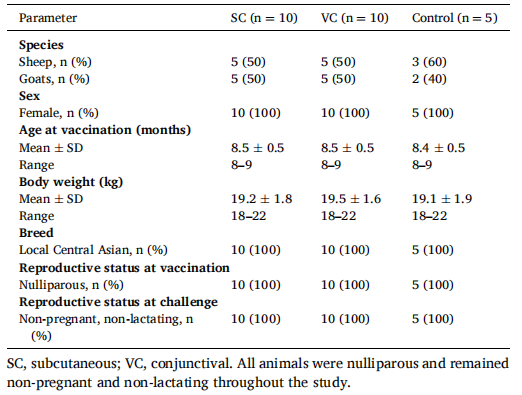
Table 2 Individual ELISA S/P% values in animals vaccinated subcutaneously (SC) or conjunctivally (VC) with Brucella melitensis Rev.1 from baseline to 15 months after vaccination
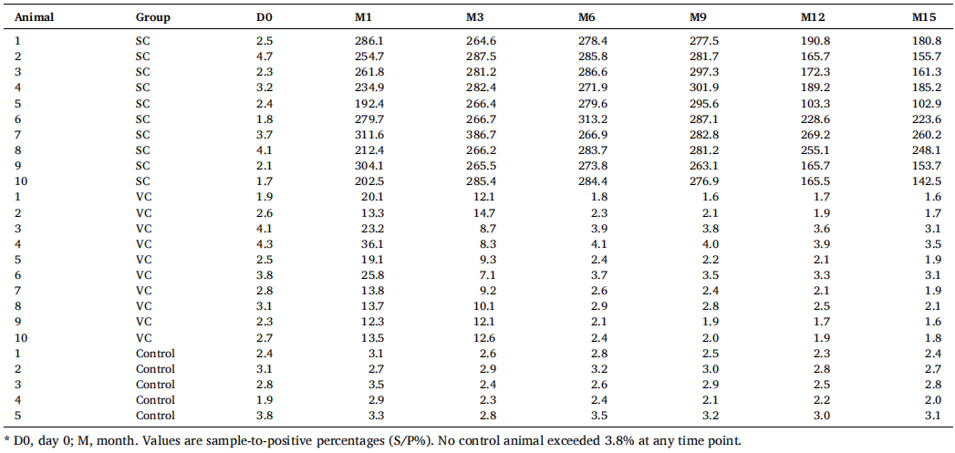
Table 3 Summary of ELISA S/P% values by group and time point, with between-group comparisons
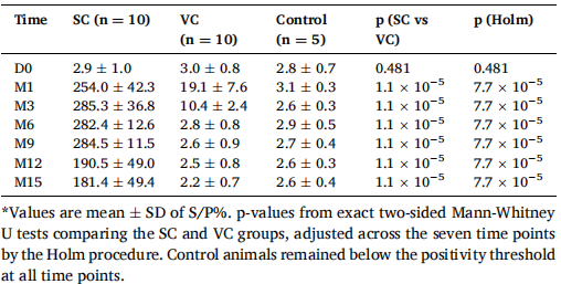
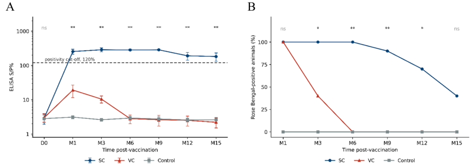
Figure 1 Serological responses following subcutaneous (SC) and conjunctival (VC) vaccination with Brucella melitensis Rev.1. (A) ELISA antibody kinetics: group means ± SD of the sample-to-positive percentage (S/P%), plotted on a logarithmic scale; the dashed line indicates the manufacturer's positivity cut-off of 120%. (B) Percentage of Rose Bengal-positive animals. Asterisks denote the Holm-adjusted significance of the SC versus VC comparison: *p < 0.05, **p < 0.001; ns, not significant
Table 4 Rose Bengal test results in animals vaccinated subcutaneously or conjunctivally with Brucella melitensis Rev.1 during the post-vaccination follow-up period
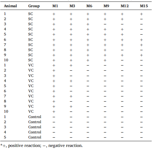
Table 5 Proportion of Rose Bengal-positive animals by group and time point, with between-group comparison
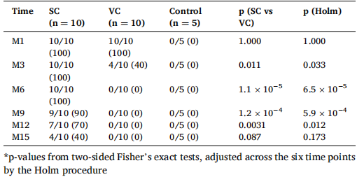
Table 6 Distribution of Brucella recovery by organ after subcutaneous vaccination (SC, n = 10)
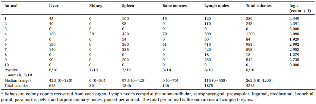
Table 7 Distribution of Brucella recovery by organ after conjunctival vaccination (VC, n = 10)
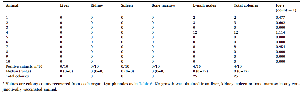
Table 8 Distribution of Brucella recovery by organ in the non-vaccinated control group (n = 5)
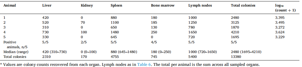
Table 9 Summary of bacterial recovery and organ colonisation by group, with 95% confidence intervals and adjusted p-values
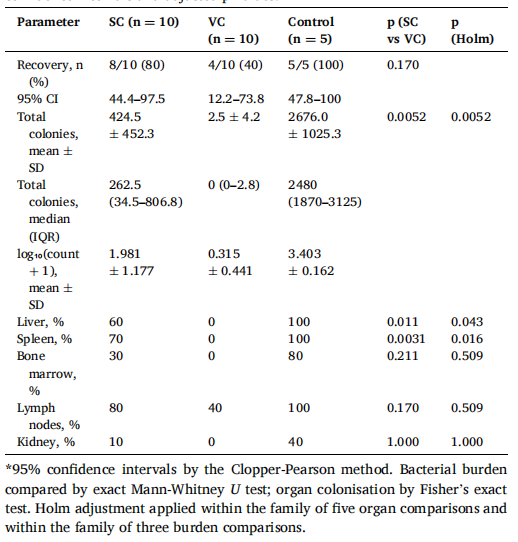
Table 10 Identification of Brucella isolates recovered after challenge
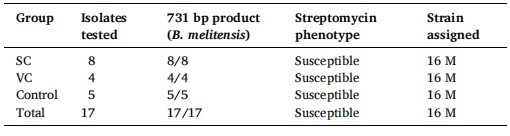
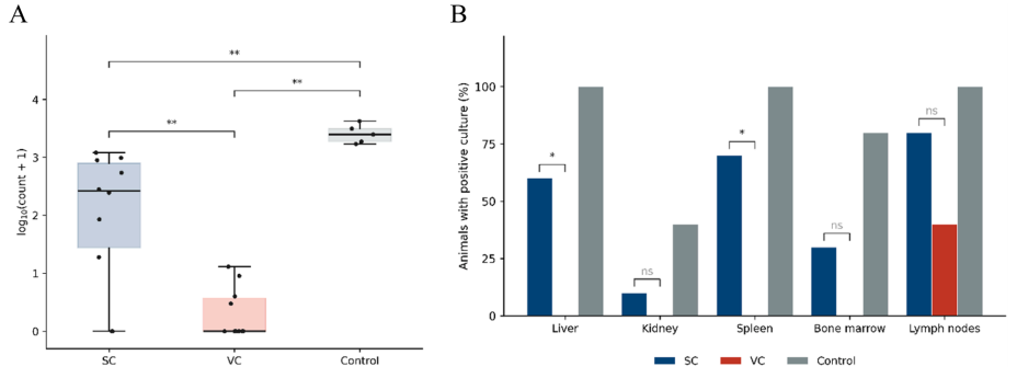
Figure 2 Bacterial burden and organ colonisation after experimental challenge. (A) Total bacterial burden per animal, expressed as log₁₀(count + 1); boxes show the interquartile range, horizontal lines the median, whiskers the range, and points individual animals. (B) Percentage of animals with a positive culture from each organ. Asterisks denote Holm-adjusted comparisons: *p < 0.05, **p < 0.01; ns, not significant
附件:
 返回列表